
Microplastics 101:
From Synthetic Beads to Real-World Particles
Microplastics are everywhere — but how we study them matters
Micro- and nanoplastics are now detected in air, water, food, and human tissues. As interest in their potential impact on human health grows, so does the need for reliable experimental systems. However, there is a growing gap between what exists in the real world and what is used in the lab.
What most studies use: synthetic plastic beads
Most microplastic and nanoplastic studies rely on commercially available polystyrene nanospheres.
These materials are widely used because they are:
uniform in size
easy to detect
experimentally convenient
They were originally developed for controlled assays or as size standards, not to represent real-world exposure.

Where the mismatch begins
Real-world micro- and nanoplastics are:
irregular fragments, not perfect beads
polydisperse, spanning a wide range of sizes
chemically heterogeneous, reflecting industrial materials and degradation
In contrast, synthetic nanospheres are:
perfectly spherical
monodisperse
often surface-functionalized (engineered with chemical groups or charges that alter how they interact with proteins and cells)
Why this matters in biological studies
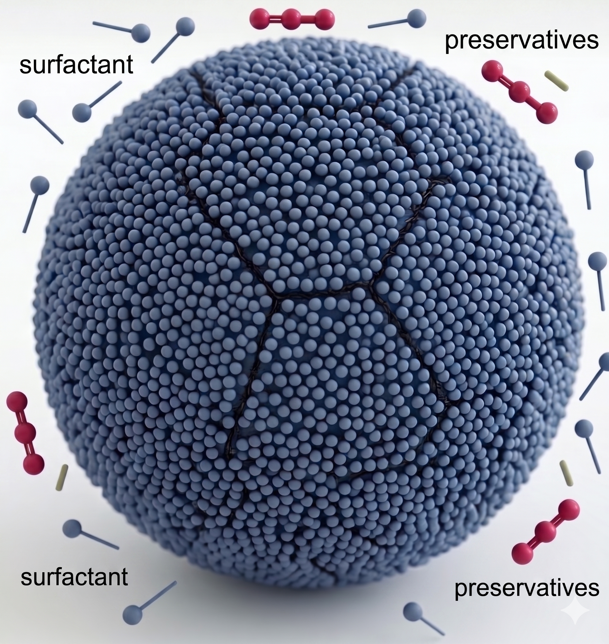
Many synthetic nanosphere preparations are not simply “plastic particles.”
They are formulated with:
surfactants in solution
surfactant layers on the particle surface (total surface area: 57 m² in 1g of 100 nm spheres)
cytotoxic preservatives
These components can:
disrupt membranes
alter uptake pathways
dominate observed biological effects
What appears to be a plastic exposure experiment may, in part, be a surfactant exposure experiment.
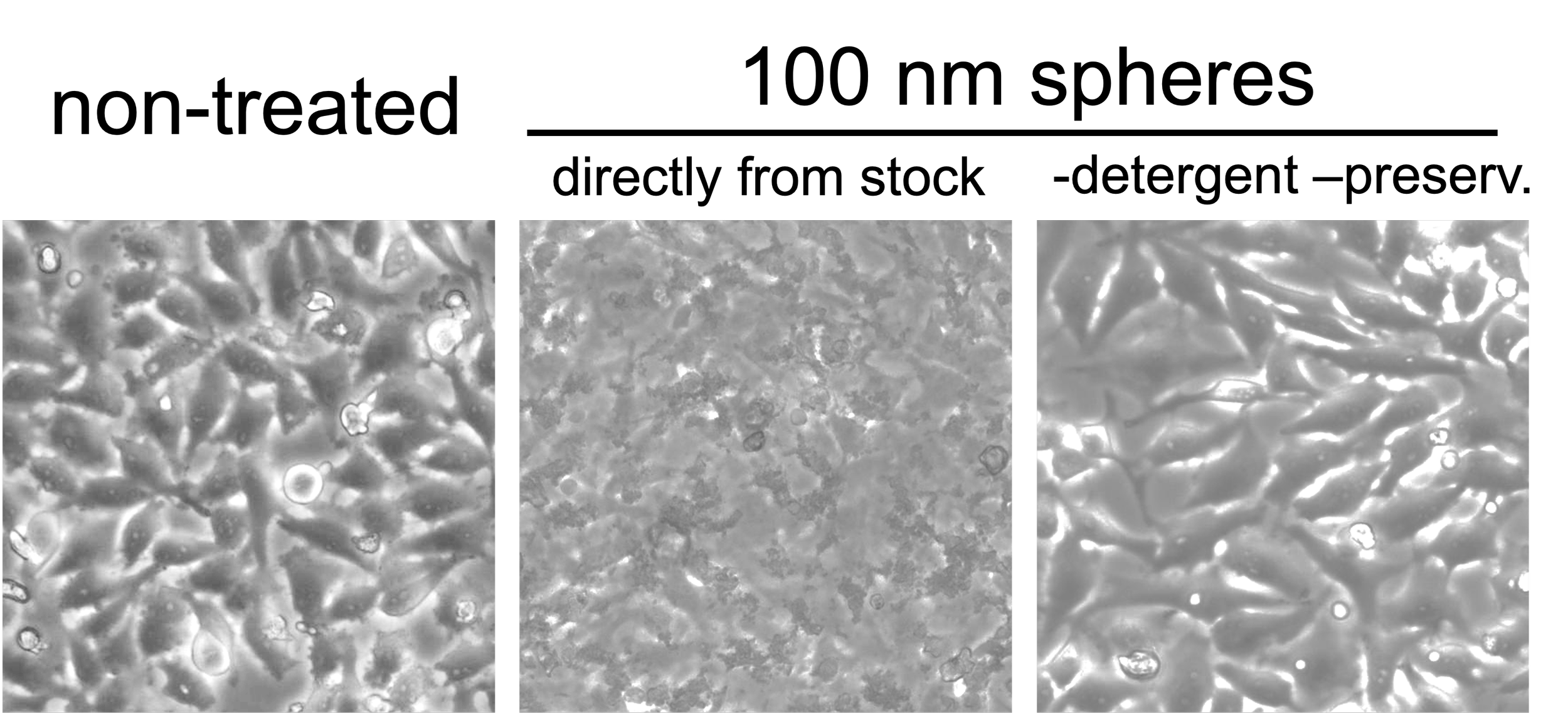
Case observation: formulation can dominate biology
In our experiments, HeLa cells exposed to polystyrene nanospheres (from a major commercial vendor) at the upper end of commonly used conditions (100 nm, 500 µg/mL) showed rapid cell lysis within 1 hour in the absence of serum.
In contrast, when surfactants were removed and particles were stabilized with minimal BSA,
this effect was not observed under the same conditions.
This suggests that formulation—not just the plastic itself—can drive observed cytotoxicity.

VS

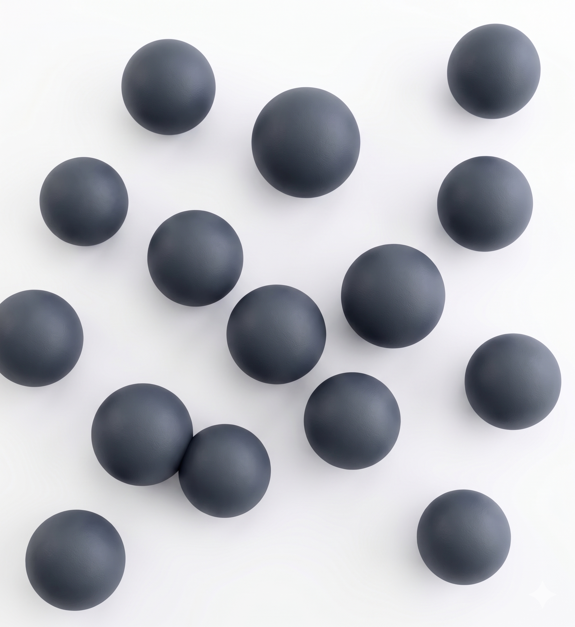
Spheres vs. Fragments
Nanoparticles are similar in size to large biological structures — shape matters
Fragments present greater surface area (up to ~10×) than spheres
Increased surface area enables more molecular and cellular interactions
Irregular geometry promotes intracellular aggregation and persistence

Toward more biologically relevant models
When synthetic nanospheres are used as stand-ins for real nanoplastics, experimental outcomes may reflect:
surfactant effects
surface-driven artifacts
idealized particle geometry
rather than true particle–biology interactions.
To better align experiments with real-world exposure, there is increasing interest in materials that:
reflect realistic particle morphology
preserve native polymer properties
minimize formulation-driven artifacts
Mechanically generated plastic fragments represent one approach to bridging this gap—improving reproducibility and enabling more biologically meaningful results.